


Before we get into the details about gas exchange, surface area to volume ratio oxygen and carbon dioxide concentration, lets look at the actual structure of the human respiratory system.
The first thing you will notice is that both the nose and mouth link to the Trachea.
This means air taken in through the mouth goes to the same place as air taken through the nose.

Gas Exchange

However it is the preferable that air is taken in through the nose.
As air enters the nose it can be filtered, moistened and warmed through the nasal passages.
Air taken in is pushed to the back of the throat (pharynx).
The Trachea is a long tube which travels from the pharynx to the lungs.
Its held open with ‘C’ shaped rings of cartilage. This prevents the trachea from collapsing when there is no air flow.
The trachea and bronchi are lined with ciliated cells which produce mucus, but more on that a bit later


At the top of trachea is the Larynx (voice box). This is where the vocal cords are located.
On top of the larynx is the epiglottis. When this cartilage flap is closed, it stops food and saliva from entering your lungs.
The vocal cords allow us to change our pitch and tone. When we whisper, yell, talk in a deep voice or high pitch voice, we use our vocal cords.
As you can see from the video to the left, Your vocal cords allow you to change your pitch without inhibiting air flow.
In men, you may see a bump on their throat. This is referred to as an ‘Adam’s apple’ and although both men and women have them, its much more visible on men.
All it is, is a chunky bone of cartilage that’s wrapped around the larynx. The technical name is Thyroid cartilage.

Testosterone causes cartilage to grow to twice its original size. Its main function is to protect the larynx, and as a bi-
You can see to the left, both a males and females ‘Adam’s apple’

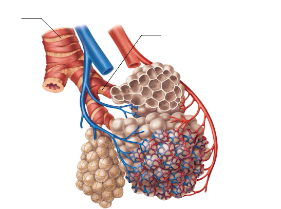
Moving down the trachea, it eventually splits forming two tubes called Bronchi.
Within the Trachea and Bronchi are ciliated cells (click here for information on cilia) which produce mucus.
As well as ciliated cells, Goblet cells also produce special mucus to trap dust and pathogens.
The mucus traps dust and bacteria brought in during breathing. The cilia then push them back up towards the mouth, so it can be swallowed.

If we have a look at the lungs we can see a difference between the right and left.
The right lung has 3 lobes, where as the left only has 2 lobes. Another thing you will notice is dip in the left lung.
This is called the Cardiac notch. The heart sits just behind here.

The Bronchi continue to branch into smaller tubes called Bronchioles.
They eventually lead into small air sacs called Alveoli. This is where the majority of gas diffusion occurs. Our lungs contain over 300 million alveoli.
Capillaries are wrapped around the Alveoli. This allows diffusion of gasses through the blood stream. We will talk more about this later on.
By constantly branching out we increase the SA:V ratio. Our lungs have roughly the same SA of a tennis court.
When we talk about the respiratory system, they first thing you might think about is the amount of oxygen in your body. However the body actually measures the amount of carbon dioxide in the body not oxygen. There are small carotid bodies (receptors) located on carotid arteries, which detect the amount of Carbon dioxide in the blood.
If the concentration of Carbon dioxide is too high and a message is sent causing the Diaphragm to contract and the ribs to expand. This increase the amount of oxygen in the blood and decrease the concentration of Carbon dioxide.

Now we know the pathway into our lungs, how do we actually breath
How do our Lungs actually expand?
Click here to learn more about Lung ventilation and Tidal Volume
Now that you know the basic components of the human respiratory system. Lets take a look how oxygen gets from your lungs to all your body cells.
Click here for information on alveoli
Complete the questions bellow on the respiratory system.
1. Write the following in order as air enters the mouth
Vocal cords
Epiglotis
Alveoli
Bronchus
Pharynx
2. Explain why it is beneficial to breath in through the nasal passage.
3. Explain the function and structure of the alveoli. 2 marks
4. Explain the structure of the trachea. 2 marks
Research
For this part you will need to research the following to diagnose your patient.
Old man Jackson has recently walked into your clinic. For the past 3 days he has been complaining of chest pain when breathing and coughing. You take his temperature and notice he has a fever. Even though his temperature is high, he explains that he has has shaking chills.
As you continue to ask about any other changes and symptoms of a possible infection he explains that he has also had nausea, vomiting, shortness of breath and Fatigue.
In your books, write out the symptoms Old man Jackson exhibits
5. Based on the evidence above what could he have?
(hint, this is a respiratory disease starting with p)
6. From your diagnosis, you have discovered that this is a bacterial infection, which caused the disease. What is your course of action as a doctor?
7. Based on the information above, what defensive measures did the bacteria avoid in the respiratory system to cause an infection? (3 marks)
8. Explain how an asthma attack affects the lungs. Use the following words in your answer: Bronchi, trachea, coughing, cartilage, inflamed and mucus. 7 marks.
9. Draw the diagram in to your book and fill in the labels.
We know the respiratory system is made up of tubes, lungs, defence cells and bones, but there are also muscles which you may not know about.


